Vista lateral del pie derecho mostrando los huesos del pie y el tarso. Imagen por Lecturio. Articulaciones del pie. Las articulaciones del pie, de proximal a distal, incluyen las siguientes articulaciones. Articulación subastragalina o astragalocalcánea: Tipo: articulación sinovial o articulación sinovial plana. (MCL), allowing for release. Depicted are the initial (left) and final (right) positions of the hand showing the lateral rotation of the wrist necessary for needle translation in the pie crust maneuver during release of a right knee MCL. Figure 5. The medial joint space of the knee should be visualized simultaneously during the performance of this

Los huesos del pie Todo lo que necesitas saber Pies Sanos, Podólogo Valencia

Bones of the Lower Limb Anatomy and Physiology I
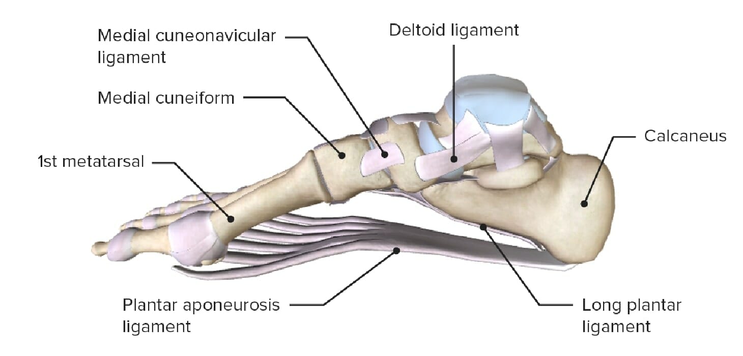
Pie Anatomía Concise Medical Knowledge
:watermark(/images/watermark_5000_10percent.png,0,0,0):watermark(/images/logo_url.png,-10,-10,0):format(jpeg)/images/overview_image/2142/JvWdPTYKrY0Ym4pUsX0Vow_ankle-joint-medial_spanish.jpg)
Tobillo y pie Huesos, músculos, articulaciones Kenhub
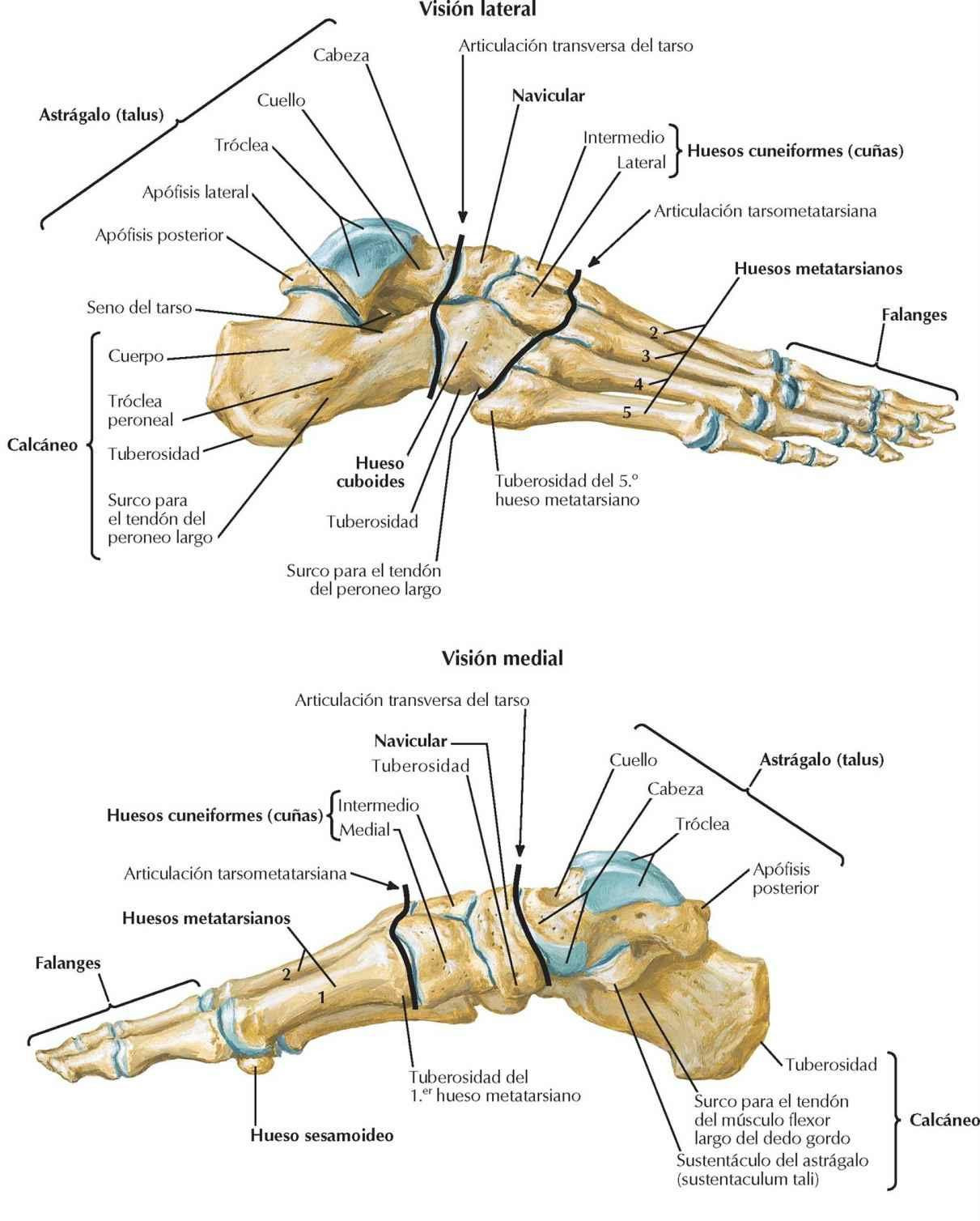
Huesos Del Pie Y Tobillo

Huesos del pie
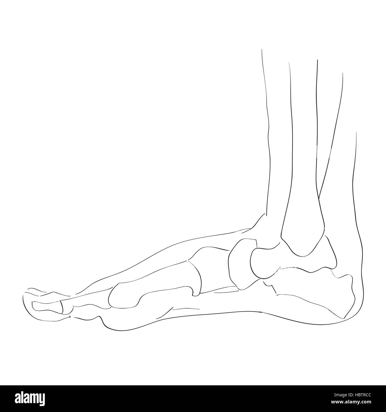
Huesos del pie fotografías e imágenes de alta resolución Alamy
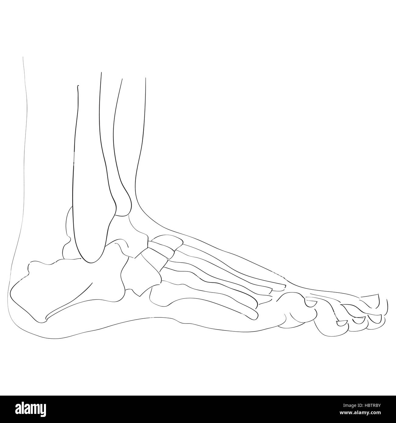
Hueso De La Vista Lateral Del Pie Fotos e Imágenes de stock Alamy
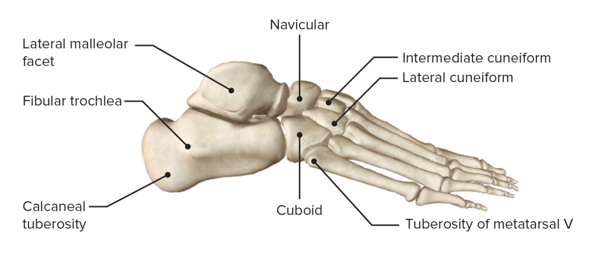
Pie Anatomía Concise Medical Knowledge

Los 26 huesos del pie humano (y sus funciones)

Huesos del pie ¿Cuántos son? Funciones, anatomía, partes y mucho más
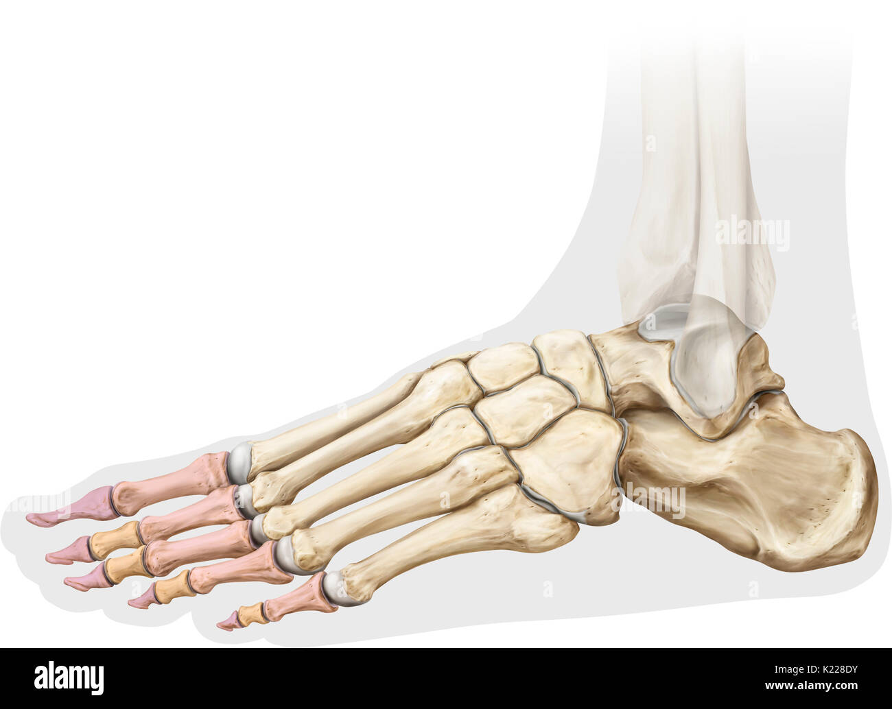
Hueso De La Vista Lateral Del Pie Fotos e Imágenes de stock Alamy
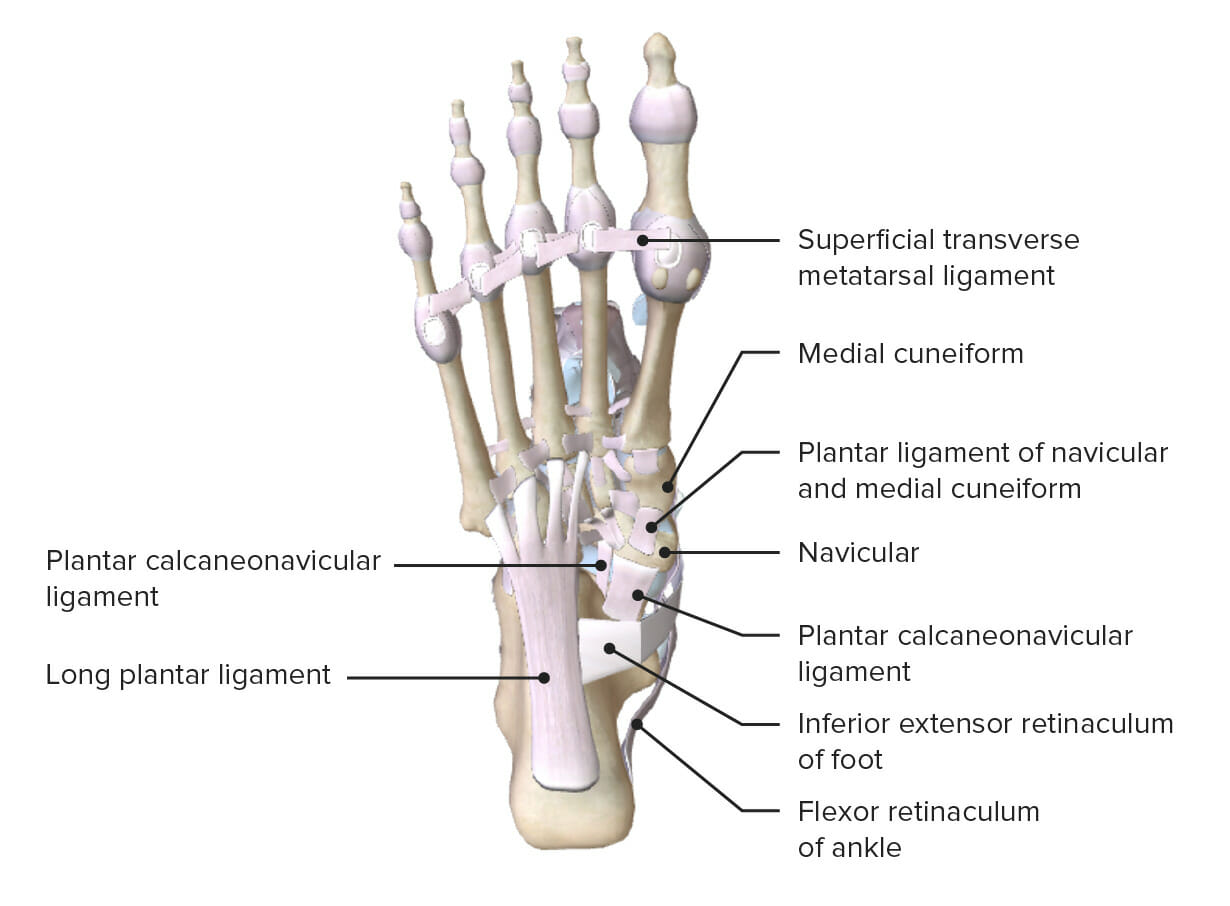
Pie Anatomía Concise Medical Knowledge

Huesos del Pie Apuntes médicos Apuntes digitales uDocz
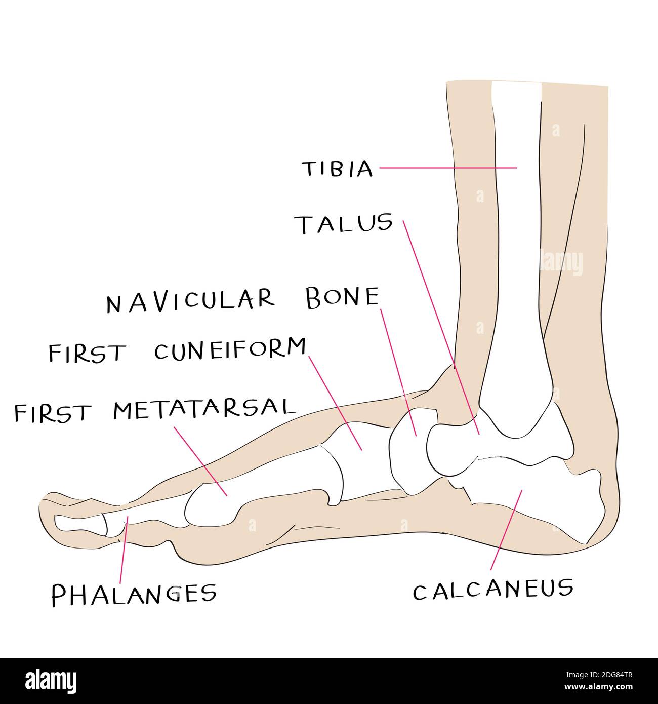
Hueso De La Vista Lateral Del Pie Fotos e Imágenes de stock Alamy

Estructura Anatómica De Los Huesos Del Pie Humano. Stock de ilustración Ilustración de
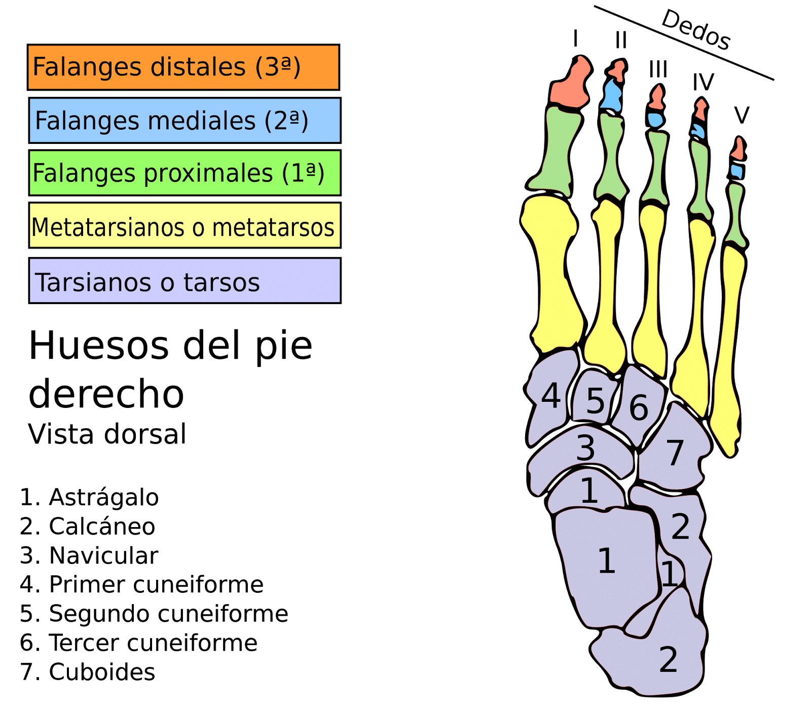
Músculos del pie anatomía, funciones, origen e inserción y más
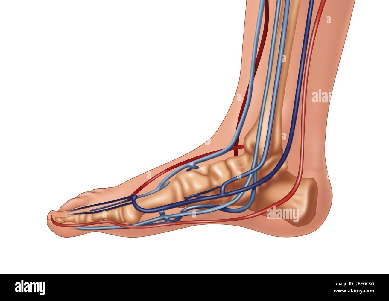
Anatomía del pie, Ilustración Fotografía de stock Alamy

Huesos del pie Vista Lateral 3D CreativeMed
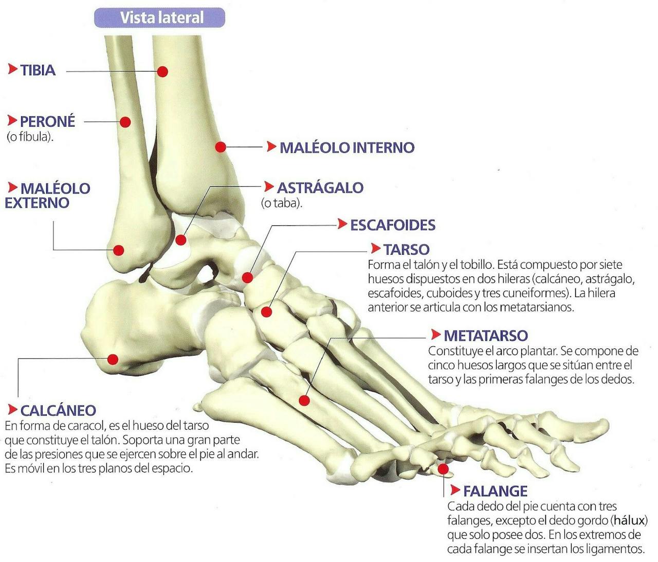
Los 14 tipos de pies (y cómo identificar el tuyo)
The piece of pie sign refers to an abnormal triangular appearance of the lunate on a PA image of the wrist representing either lunate dislocation or perilunate dislocation 1,2. A lateral image will help differentiate whether there is lunate or perilunate dislocation, with lunate dislocation demonstrating a spilled teacup sign. This is largely.. Estiramiento de la banda iliotibial de pie: Cruce la pierna sana por delante de la afectada y agáchese sin doblar las rodillas, intentando tocar la punta de.